Close


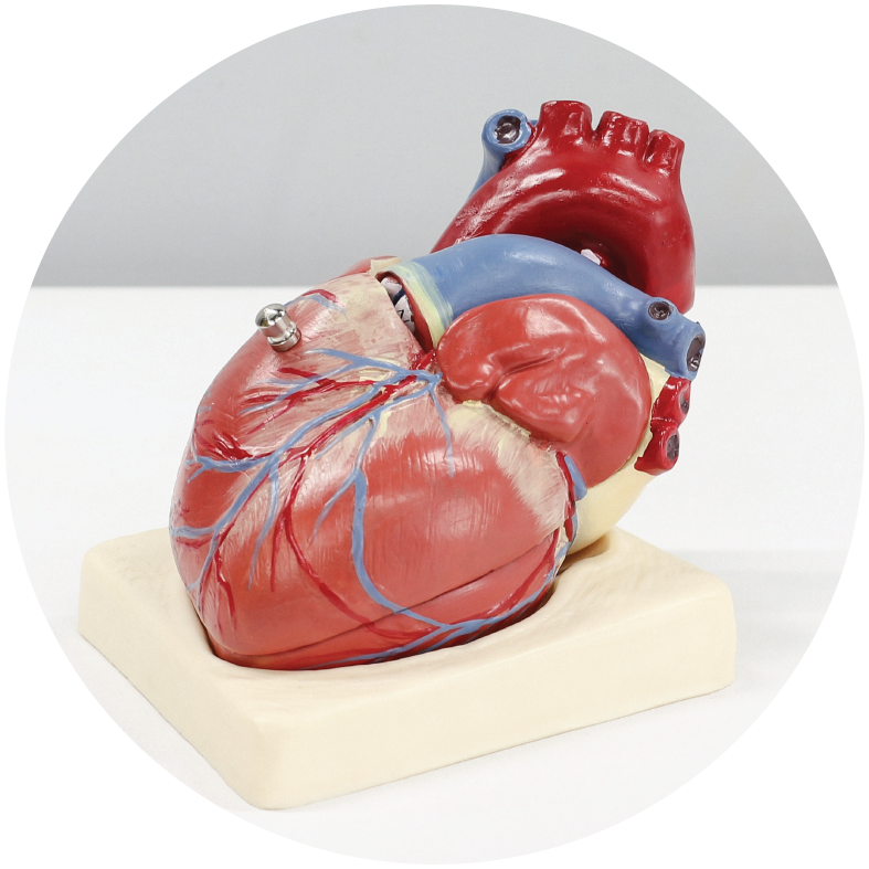
The cardiovascular system consists of the heart and the vessels. The heart is made up of an extremely powerful muscle, the myocardium, which is the motor of blood circulation. By acting as a pump, the heart allows blood to move through the vessels.
The heart is divided into four chambers, two on the right and two on the left. The heart and vessels together form the cardiovascular system, also known as the circulatory system.
The path of the blood
The blood flows along an unchanging path regulated by the heart’s contractions and a set of one-way valves that prevent backward flow.
The starting point of the circuit is the left ventricle, which is the most muscular part of the heart.
During the contraction of the left ventricle, the blood is ejected into the arterial network and then successively reaches the capillary and venous networks. During this journey, the blood gradually gives up its oxygen and the sugar necessary for the proper functioning of the organs and recovers the various waste products produced by the cells of our body, including carbon dioxide.
The venous network is connected to one of the two right chambers of the heart, the atrium. The blood that enters the atrium is then propelled to the second right chamber, the ventricle. When the right ventricle contracts, it expels blood to the lungs, where the blood is freed from the carbon dioxide it has stored and recharged with oxygen. This reoxygenated blood is redirected to the left atrium and from there is propelled into the left ventricle. This cycle is repeated with each heartbeat.
The circulatory system contains about 5 litres of blood. Each heartbeat mobilises about 60 ml of blood.
Heartbeats
Such precision mechanics are regulated by an automatic pacing system in the wall of the right atrium, so that the heart has its own pacemaker.
The stimulation centre is called the sinus node. This is a group of cells that have the property of contracting automatically in a rhythmic way and triggering the contraction of all the cells of the myocardium from one to the other according to a very precise circuit. The contraction first involves the two atria, then the two ventricles. The two atria contract simultaneously, as do the two ventricles. The rate of contraction of the cells of the sinus node corresponds to the heart rate, which varies according to the needs of the body. At rest, the heart rate is around 50 to 70 beats per minute. During exercise, it can exceed 200.
Coronary circulation
As the heart is a muscle, it also needs oxygen and glucose. These elements are supplied by a special arterial network, the coronary network, which is located on the surface of the heart and which has the particularity of having a blood circulation that takes place during the period when the heart chambers are not contracting, i.e. diastole.
Cardiovascular abnormalities
Each of the components of the system may function abnormally, either in isolation or more globally. The most common heart problems are :